What is larval settlement and metamorphosis?
Norman J. Blake, in Developments in Aquaculture and Fisheries Science, 2016 Larval settlement and metamorphosis represent a period of high mortality since it is a period of their life history in which larvae undergo behavioural changes associated with their search for an appropriate substrate upon which to settle.
What is larval settlement in corals?
Larval settlement. Settlement is a crucial step in the life history of a coral. It is about time to switch from a mobile planktonic phase to a sessile one. Therefore, the choice of a suitable settlement place is important and, moreover, a conscious decision.
How do microorganism biofilms affect larval settlement in invertebrates?
Chemical cues associated with microorganism biofilms also contribute to larval settlement for a wide range of sessile invertebrates, including sponges and corals [ 20, 21, 32 ]. Consistent with settlement responses of coral larvae to CCA, settlement to biofilms can also be variable for some species [ 33 ].
Does surface microtopography play a role in larval settlement for some larvae?
The notable outcome of this study was that surface microtopography plays a substantial role in settlement for some larvae, in part supporting previous studies on corals and sponges that have established the importance of surface topography in larval recruitment [ 37 – 40 ].

Molecular mechanisms of Platynereis larval settlement
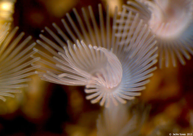
Some marine zooplankton are only temporary residents of the plankton, representing the larval stages of marine invertebrates such as corals, worms, snails, clams and crabs. After a period in the open water, these larvae must settle to the ocean floor where they will live out their adult lives.
MIP peptide treatment triggers Platynereis larval settlement
MIP treatment triggers Platynereis larval settlement in 2 day larvae. The video shows a timelapse recording of untreated larvae (left) and larvae in the presence of 5 µM MIP. In the right side larval tracks are marked in white, and the current larval position in red.
Chemosensory neurons in the larval apical organ
Chemosensory cells in the 2 day Platynereis larvae. These cells can be stained by dye-filling with Mitotracker. Some of these cells express the neuropeptide MIP. The image is color coded for depth. Scale bar 20 µM.
Expression of MIP and its receptor in the apical organ
The neuropeptide MIP (red) is expressed in chemosensory-neurosecretory cells in the apical organ of 2 day Platynereis. Immunostaining against acetylated tubulin (white) highlights the cila and axonal scaffold of the larva.
Serial TEM reconstruction of a pair of MIP-expressing chemosensory-neurosecretory neurons
Transmission electron microscopy reconstruction of a pair of MIP-expressing chemosensory-neurosecretory cells. The cells have long branching microvilli and are packed with many dense core vesicles, indicating that they have both chemosensory and neurosecretory character.
How to do larval settlement?
Larval settlement assays for each species were undertaken by pipetting 20 larvae into 20 mL glass scintillation vials holding 15 mL of 10 μm filtered sea water (FSW), placing the artificial settlement tile over the opening of the scintillation vial, and carefully inverting the vial so that larvae were in direct contact with the test surface. Vials were then secured to surfaces with rubber bands and maintained in temperature controlled rooms with 12:12 photo period cycles. A total of 10 replicate tiles for each of the surface sizes were used for each species. Larval metamorphosis was scored every six hours using an inverted microscope. Larval metamorphosis was scored in both holes and on the flat surface between holes for all surface treatments, and the flat surface of controls. While we acknowledge there are differences between the terms of “settlement” and “metamorphosis” [ 21] for clarity we use the term “settlement” as a descriptor of the process of larval settlement through to larval metamorphosis from this point on.
How does microtopography affect larval settlement?
Compared to chemical cues, the effect of physical microtopography on larval settlement is less well studied. Surface structure, at topographical scales that approximate larval sizes (i.e. < 1 mm), is important for some temperate invertebrate larvae, such as barnacles [ 34 – 36 ]. Surfaces structured with crevices of several millimetres have been used in coral and sponge recruitment studies [ 37 – 41] but these studies demonstrated settlement to surface topography that were considerably larger than larval/propagule dimensions. There is considerable knowledge on the role of microtopography in the fields of marine biofouling and bio-mimicry [ 42, 43] with a predominant focus on determining methods to impede larval settlement. One of the key findings from this research is that settlement and adhesion of marine invertebrate larvae can be influenced by the complexity of surface microtopography, a concept developed through “Attachment Point Theory” [ 44 ]. If microtopography provides surfaces with texture that closely match larval dimensions, then settlement is increased by increasing the available points of attachment for larvae or propagules [ 45, 46 ]. Conversely, surfaces that are smaller than larval dimensions minimise the number of attachment points and often reduce settlement success [ 44] and the strength of adhesion [ 47, 48 ].
What are the chemical cues that signal habitat and illicit larval settlement?
Chemical (environmental) cues that signal habitat and illicit larval settlement are a common denominator for a wide range of sessile marine taxa with settlement initiated in response to conspecifics [ 17 ], host organisms [ 18] and microorganism biofilms [ 19 – 21 ]. Two of the most interesting chemical cues implicated in the settlement of coral reef invertebrates involve crustose coralline algae (CCA) and microbial biofilms.
Why is larva size important?
Larval size is important in interpreting the effect that specific sizes of microtopography have on larval settlement choices. Therefore, 50 larvae from each of the five species were collected at spawning (sponges), or just prior to use in settlement assays (corals), and fixed in 2.5% glutaraldehyde to enable measurements of larval width and length.
Where are C. crassa and A. millepora found?
A. millepora and C. crassa are locally abundant corals found throughout the shallow waters of the Great Barrier Reef. A. millepora is hermaphroditic and C. crassa is gonochoric. Both species are broadcast-spawning corals with non-feeding larvae that lack zooxanthellae when released.
What is habitat selection?
Habitat selection can revolve around complex behaviours with some taxa demonstrating defined and innate behaviours that optimise selection [ 5, 6 ] . This is often displayed for breeding [ 7 ], with marine turtles returning to natal beaches to lay eggs [ 8 ], brood parasitism in birds [ 9, 10 ], and insects using chemical cues to oviposit eggs in habitats that minimise predatory risks, or optimise food resources for emerging larvae [ 11] being among the many examples identifying the use of behaviour to provide optimal habitats for offspring.
Can larval settlement be driven by microtopography?
In conclusion, this study has established that larval settlement can be driven by physical (microtopography) settlement cues. It also questions the sole reliance on chemical cues for larvae to settle. While the long term survival of recruits was not a focus of this study, future work incorporating both chemical and physical cues, coupled with data on post settlement survival will provide an increased understanding of the dynamics that drive larval settlement and recruitment.
Acknowledgements
This research is funded in part by the Gordon and Betty Moore Foundation through Grant GBMF5009 and the Office of Naval Research grant no. N00014-08-1-2658 to MGH. The authors would also like thank Mrs. Tina M.
Author information
University of Hawaii, Kewalo Marine Laboratory, Honolulu, 96813, United States
Rights and permissions
This work is licensed under a Creative Commons Attribution 4.0 International License.
Comments
By submitting a comment you agree to abide by our Terms and Community Guidelines. If you find something abusive or that does not comply with our terms or guidelines please flag it as inappropriate.
